Share
Modern Applications of Machine Learning in Radiology

- BLOG
- Artificial Intelligence
- March 3, 2026
Radiology rooms process thousands of images daily, yet even expert eyes can miss subtle patterns hidden in pixels. That is where machine learning in radiology steps in, not as a replacement, but as a second set of analytical eyes trained on massive datasets.
As imaging volumes rise and cases grow more complex, relying only on manual interpretation becomes increasingly demanding. Radiology machine learning models analyze texture, structure, and density at scale, supporting consistency while helping specialists focus on higher-level clinical reasoning.
Against this backdrop, understanding how ML in radiology truly functions becomes essential. In the sections ahead, we examine its real applications, data foundations, evaluation standards, risks, and what its growing role means for modern diagnostic practice.
Contents
- 1 What is Machine Learning in Radiology?
- 2 Why Radiology Is a Strong Fit for Machine Learning
- 3 Major Applications of Machine Learning in Radiology
- 4 Deploy Production-Ready Radiology ML Systems.
- 5 What Data Radiology ML Models Actually Need
- 6 How to Evaluate a Machine Learning Model in Radiology
- 7 Regulatory and Compliance Considerations for ML in Radiology
- 8 Risks and Limitations of Machine Learning in Radiology
- 9 Commercial Radiology AI Tools vs Custom Machine Learning Development
- 10 Working with the Right ML Partner in Radiology
- 10.1 We design models around real radiology decisions
- 10.2 We engineer for PACS-driven imaging environments
- 10.3 We build models trained for radiology-specific data variability
- 10.4 We implement lifecycle monitoring for sustained clinical reliability
- 10.5 We deliver long-term strategic capability, not one-time models
- 11 Future Directions of Machine Learning in Radiology
- 12 Deploy Production-Ready Radiology ML Systems.
- 13 Conclusion
- 14 Frequently Asked Question
What is Machine Learning in Radiology?
Machine learning in radiology uses algorithms trained on large medical image datasets, including CT, MRI, X-ray, and ultrasound scans, to identify patterns and assist diagnosis. These models can detect abnormalities, classify findings, measure structures, and assist with interpretation.
Unlike rule-based software, machine learning systems improve through exposure to data. In radiology, this commonly involves deep learning in medical image analysis, along with natural language processing to structure radiology reports.
Overall, machine learning serves as a decision-support tool that enhances efficiency and consistency while keeping radiologists in control of clinical judgment.
Why Radiology Is a Strong Fit for Machine Learning
 The importance of machine learning in radiology becomes clear when examining the structural nature of the specialty.
Radiology produces high-volume, data-rich, visually standardized outputs that align closely with how modern machine learning models are trained, validated, and deployed in clinical environments.
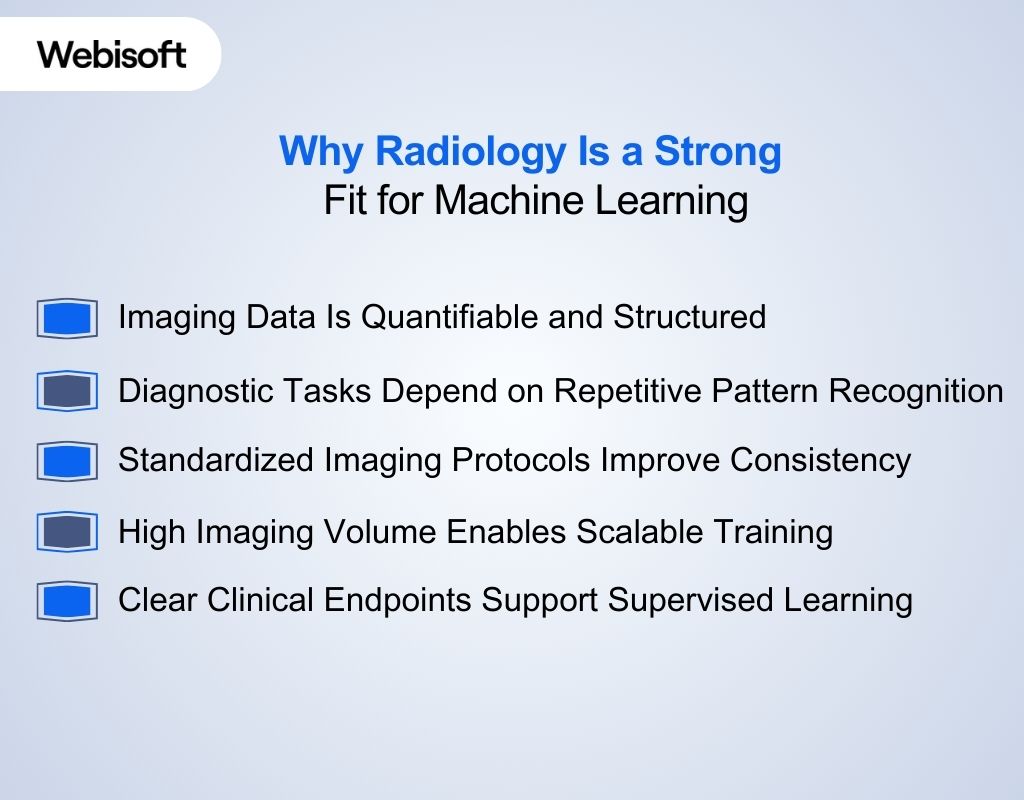
Imaging Data Is Quantifiable and Structured
Radiology generates pixel-level numerical data rather than narrative observations. CT, MRI, and X-ray scans consist of structured intensity matrices, making them computationally suitable for supervised learning. Unlike specialties that depend heavily on descriptive notes, radiology data is inherently digitized and measurable.
Diagnostic Tasks Depend on Repetitive Pattern Recognition
Radiologic interpretation often involves identifying recurring visual patterns such as nodules, hemorrhage, fractures, or tissue density changes.
These tasks align well with deep learning architectures designed for spatial feature extraction and image classification.
Standardized Imaging Protocols Improve Consistency
Although scanner variability exists, imaging protocols follow defined acquisition parameters and file standards such as DICOM. This relative standardization enables multi-center data aggregation and supports more stable algorithm development.
High Imaging Volume Enables Scalable Training
Radiology departments generate thousands of studies daily. Large data availability allows models to train on diverse cases, which is important for improving performance and generalizability across patient populations.
Clear Clinical Endpoints Support Supervised Learning
Radiologic findings are frequently confirmed through biopsy results, surgical outcomes, or longitudinal follow-up. These objective endpoints provide reliable ground truth labels, which strengthen model training compared to specialties where outcomes are less clearly defined.
Major Applications of Machine Learning in Radiology
 Machine learning has moved beyond theory and now plays a practical role across core radiology tasks, improving diagnostic accuracy and efficiency through production-grade AI/ML systems.
These applications span from routine image interpretation to advanced quantitative analysis that supports clinical decisions.
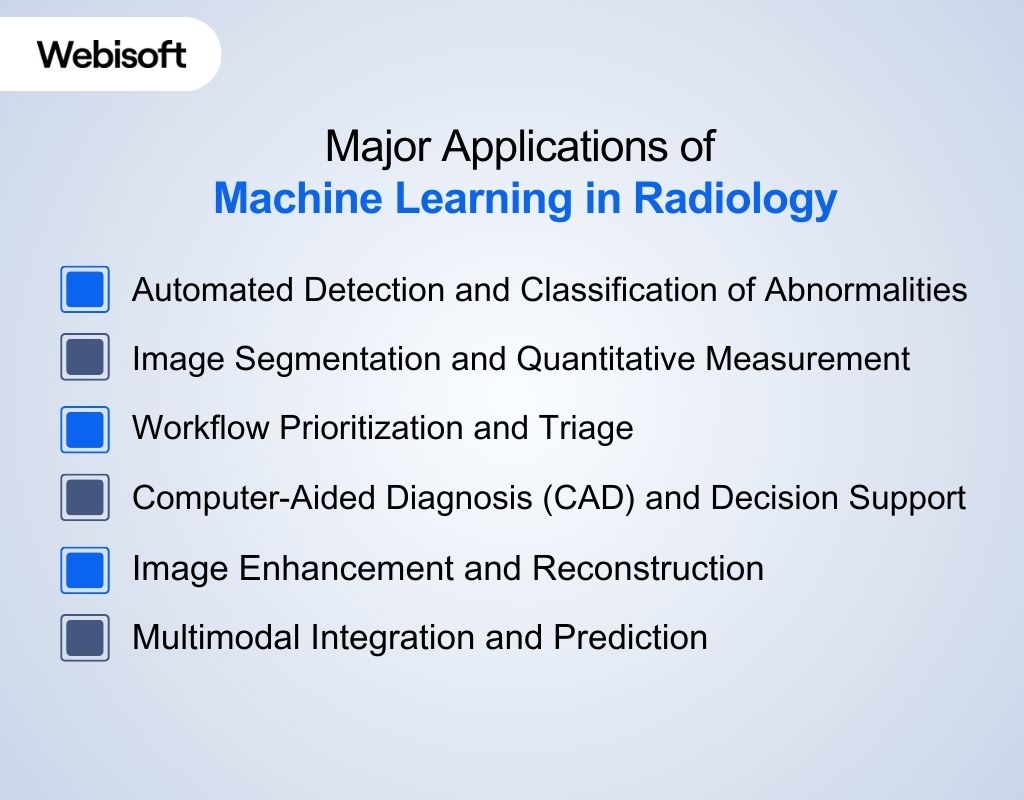
Automated Detection and Classification of Abnormalities
Machine learning models are widely used to flag suspicious findings in imaging studies by recognizing patterns that may indicate disease. This helps radiologists identify critical pathology more consistently and quickly than manual review alone.
- Detects tumors, nodules, and lesions across modalities
- Classifies disease states (e.g., benign vs malignant)
- Reduces oversight of subtle abnormalities
- Supports diagnosis across organ systems such as lungs, breasts, and brain
Image Segmentation and Quantitative Measurement
One of the foundational tasks in radiology ML is breaking down images into meaningful regions, which allows precise quantification of anatomy and pathology.
- Segments tumors or lesions for volume measurement
- Delineates organ boundaries for surgical planning
- Quantifies tissue density or disease burden over time
- Enables standardized reporting of metrics such as tumor size
Workflow Prioritization and Triage
Machine learning helps manage growing imaging workloads by identifying urgent cases that need immediate attention and improving clinical response times.
- Flags critical findings such as intracranial hemorrhage
- Prioritizes worklists so radiologists see urgent cases first
- Reduces delays in reporting time-sensitive diagnoses
Computer-Aided Diagnosis (CAD) and Decision Support
ML-powered CAD tools act as second readers, offering supportive insights without replacing human judgment. They augment radiologist confidence and help standardize interpretations.
- Provides probability scores for disease presence
- Suggests differential diagnoses
- Highlights suspicious regions for targeted review
- Assists less experienced readers with complex interpretations
Image Enhancement and Reconstruction
Machine learning improves image quality and accelerates acquisition, enabling clearer visualization with lower radiation doses and faster scanning.
- Enhances the resolution of low-dose CT scans
- Reduces noise and artifacts in MRI and PET
- Accelerates reconstruction from raw imaging data
- Improves clarity of complex structures for better interpretation
Multimodal Integration and Prediction
Advances in machine learning are enabling models that combine imaging with other clinical data to inform prognosis and treatment planning.
- Integrates imaging data with clinical records for richer insights
- Predicts disease progression or treatment response
- Supports personalized risk stratification
- Enables comprehensive decision support beyond single modalities


Deploy Production-Ready Radiology ML Systems.
Build clinically aligned machine learning with Webisoft’s engineering expertise!
What Data Radiology ML Models Actually Need
 Machine learning models in radiology depend on more than just raw images. They require well-prepared datasets with accurate labels, consistent formats, and sufficient diversity so models learn reliable patterns across patients, imaging devices, and clinical scenarios.
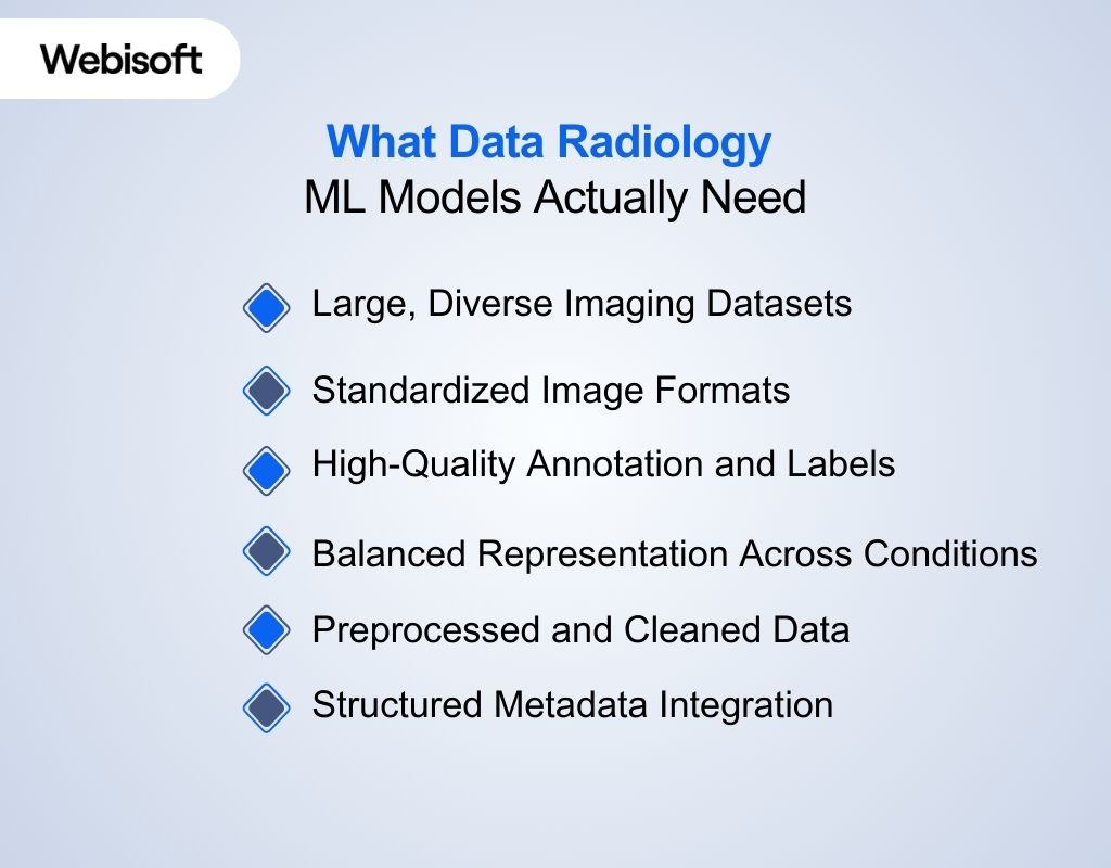
Large, Diverse Imaging Datasets
For effective model training, radiology machine learning systems need datasets with many examples covering a wide range of normal and abnormal findings. These images should come from multiple sources, imaging devices, and patient populations to help models generalize reliably.
Standardized Image Formats
Radiology datasets must be in standardized formats like DICOM, which include both pixel information and rich metadata. This standardization allows consistent image interpretation, interoperability, and integration with clinical workflows.
High-Quality Annotation and Labels
Accurate labeled data are important. Images must be annotated with clinical findings, segmentations, bounding boxes, or disease classifications so models can learn what features correspond to specific conditions. Poor labeling leads to poor model performance.
Balanced Representation Across Conditions
Datasets need balanced representation of different diseases, demographics, and imaging variations. A lack of balance can lead to biased models that perform well on some groups but poorly on others.
Preprocessed and Cleaned Data
Imaging data often requires preprocessing such as normalization, noise reduction, alignment, and resizing. Prepared data reduces technical variability and helps training focus on clinical patterns rather than artifacts.
Structured Metadata Integration
Beyond images, radiology ML models benefit from structured metadata, such as patient age, modality type, and clinical labels. So they can correlate imaging features with clinical context during training.
How to Evaluate a Machine Learning Model in Radiology
 Before machine learning systems are trusted in clinical settings, their performance must be critically examined under realistic conditions.
Understanding the role of AI in radiology requires careful validation to ensure models are accurate, reliable, and safe across diverse patient populations and imaging environments.
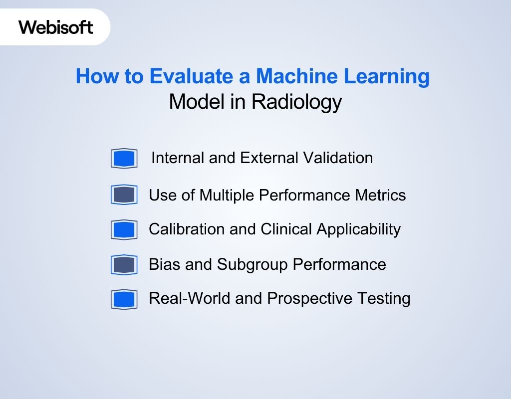
Internal and External Validation
Evaluation begins with testing on separate datasets to see if the model performs well beyond its training data. Internal validation checks performance using held-out data from the same source.
External validation tests are conducted on entirely independent data that reflects different patient populations or imaging protocols. External testing is important because models may degrade when used outside their original data environment.
Use of Multiple Performance Metrics
No single metric tells the full story. A combination of standard measures helps capture different aspects of performance:
- Sensitivity and specificity show how well the model detects true disease and avoids false alarms.
- Area under the ROC curve (AUC) summarizes discrimination ability across thresholds.
- Precision and F1 score reflect the balance between true positives and false positives in real prevalence settings.
- Dice coefficient and similar scores assess tasks like segmentation accuracy.
Using task-appropriate metrics ensures the evaluation aligns with the model’s purpose.
Calibration and Clinical Applicability
Good models should not only classify correctly but also provide output probabilities that reflect real risk. Calibration examines whether predicted probabilities match observed outcomes, which is important when a model’s output informs clinical decisions. Metrics like calibration curves or Brier scores help judge this aspect.
Bias and Subgroup Performance
Evaluation must include checks for bias and fairness. A model may perform well overall but poorly in specific demographic groups or rare conditions. Examining errors across subgroups and monitoring for data drift after deployment helps ensure equitable and robust performance.
Real-World and Prospective Testing
Finally, performance must be validated in real clinical settings through pilot deployments or retrospective case reviews that simulate actual workflow. Studies that compare model outputs with clinician interpretations on real cases provide insight into practical usefulness and limitations before full integration.
Validated radiology models still fail without strong engineering and clinical integration. See how Webisoft can integrate radiology ML into healthcare software so it fits PACS workflows, supports compliance requirements, and remains reliable in everyday clinical practice.
Regulatory and Compliance Considerations for ML in Radiology
 Machine learning systems used in radiology are regulated as medical technologies because they directly influence clinical decisions.
Regulatory oversight ensures these models are safe, clinically validated, and responsibly deployed within healthcare environments before and after approval.
Medical Device Classification and Approval Pathways
Machine learning software intended for diagnostic support is typically classified as a medical device. Regulatory requirements include:
- FDA clearance or approval through 510(k) or de novo pathways in the United States
- CE marking under the EU Medical Device Regulation (MDR)
- Clear definition of intended clinical use
- Submission of clinical performance data
- Demonstration of safety and benefit-risk balance
Approval depends on the model’s clinical role and potential patient impact.
Data Protection and Privacy Compliance
Because machine learning radiology systems rely on patient imaging data, they must comply with health data protection laws. Key obligations include:
- HIPAA compliance in the United States
- GDPR compliance in the European Union
- Secure storage and transmission of imaging data
- Proper de-identification during model training
- Controlled access to patient information
Privacy compliance must be built into both development and deployment.
Documentation, Transparency, and Traceability
Regulators require clear documentation of how the model works and where it has been validated. This includes:
- Detailed technical documentation
- Disclosure of validated patient populations
- Clear reporting of model limitations
- Defined update and version control procedures
- Audit trails for model outputs
Transparency supports accountability and clinical trust.
Post-Market Monitoring and Lifecycle Oversight
Regulatory responsibility continues after approval. Ongoing compliance requires:
- Monitoring real-world performance
- Reporting adverse events
- Managing software updates responsibly
- Re-evaluating performance after significant model changes
- Maintaining quality management systems
Machine learning systems are treated as evolving software products that require continuous oversight.
Risks and Limitations of Machine Learning in Radiology
Machine learning systems can support radiologists in meaningful ways, but they are not immune to error or constraint.
Recognizing these limitations is critical to prevent overreliance, avoid unintended harm, and maintain realistic expectations in clinical environments.
- Bias and unequal performance: If training datasets lack demographic or clinical diversity, models may perform well for some patient groups and poorly for others. This can create disparities in detection accuracy and reduce reliability in underrepresented populations.
- Generalizability challenges: A model trained on data from one hospital or scanner may lose accuracy when applied elsewhere. Differences in imaging protocols, equipment, and patient characteristics can cause performance degradation outside the original training setting.
- Sensitivity to data quality: Poor image quality, motion artifacts, or inconsistent labeling can significantly affect model output. Unlike humans, who can contextualize imperfections, algorithms may misinterpret subtle distortions as clinical findings.
- Limited clinical context awareness: Most models analyze images in isolation and do not fully incorporate patient history, laboratory results, or prior imaging. This narrow scope can restrict their ability to interpret findings holistically.
- Overfitting during development: A model may appear highly accurate during testing but fail in real-world use if it has learned dataset-specific patterns rather than true clinical features.
- Lack of interpretability: Many deep learning systems operate as black boxes. When predictions cannot be clearly explained, it becomes difficult for clinicians to assess confidence and integrate outputs responsibly.
- Automation bias risk: When AI tools are integrated into workflows, clinicians may over-trust model outputs, potentially overlooking contradictory evidence in the imaging study.
- Threshold trade-offs: Adjusting sensitivity to catch more disease often increases false positives, while tightening specificity may increase missed findings. These trade-offs must be carefully managed in clinical settings.
Commercial Radiology AI Tools vs Custom Machine Learning Development
After understanding clinical applications, evaluation standards, and regulatory requirements, the next decision centers on implementation strategy.
Healthcare organizations choose between commercial radiology AI platforms and custom machine learning solutions aligned with specific clinical and operational goals. Here’s a comparison table to differentiate:
| Aspect | Commercial Radiology AI Tools | Custom Machine Learning Development |
| Time to Deployment | Faster rollout with pre-built and pre-validated solutions | Planned development aligned with institutional roadmap and scope |
| Clinical Coverage | Optimized for common imaging tasks such as detection, triage, and segmentation | Designed to address specialized, complex, or institution-specific imaging challenges |
| System Integration | Standard integration with PACS, RIS, and reporting workflows | Engineered integration customized to existing IT architecture and workflow preferences |
| Customization Level | Feature set defined by vendor roadmap | Full flexibility in model design, feature selection, and workflow alignment |
| Data Utilization | Trained on large, multi-center datasets | Built and optimized using institution-specific imaging and performance requirements |
| Performance Optimization | Validated for broad clinical environments | Tuned for targeted clinical objectives and measurable institutional KPIs |
| Scalability Strategy | Vendor-driven product updates and feature expansion | Controlled scaling strategy aligned with long-term digital transformation plans |
Working with the Right ML Partner in Radiology
 After comparing commercial tools with custom development, the next question is who can deliver reliably within clinical constraints.
Webisoft helps you turn radiology ML plans into production systems that fit real workflows, stay stable over time, and support measurable outcomes.
We design models around real radiology decisions
Machine learning in radiology must support specific diagnostic actions, not abstract predictions. We begin by mapping the exact clinical task your model will assist.
- Define the imaging modality and pathology scope
- Align model outputs with radiologist interpretation needs
- Establish measurable diagnostic performance targets
- Build models that support decision-making, not replace it
We engineer for PACS-driven imaging environments
Radiology ML must function inside imaging ecosystems. We develop solutions that operate within real-world clinical systems.
- Integration planning with PACS and RIS workflows
- Image ingestion pipelines optimized for DICOM data
- Compatibility with structured reporting environments
- Minimal disruption to radiologist reading patterns
We build models trained for radiology-specific data variability
Medical imaging data varies across scanners, protocols, and institutions. Our approach accounts for this variability during training and validation.
- Dataset preparation customized to modality differences
- Cross-environment validation strategies
- Performance monitoring aligned with radiology use cases
- Model refinement based on institutional imaging patterns
We implement lifecycle monitoring for sustained clinical reliability
Machine learning in radiology is not static. Imaging protocols evolve, patient demographics shift, and performance can drift over time.
- Continuous performance monitoring
- Structured retraining workflows
- Drift detection planning
- Governance processes for model updates
We deliver long-term strategic capability, not one-time models
Radiology departments increasingly view machine learning as a core capability. We help you build internal systems that scale with clinical growth.
- Scalable ML architecture
- Roadmap planning for future radiology AI initiatives
- Ongoing technical partnership
- Support for expanding imaging modalities
Radiology ML initiatives succeed when clinical goals, imaging infrastructure, and long-term performance strategy are engineered together. Let’s move your machine learning in radiology project from concept to production, connect with us and start building it the right way.
Future Directions of Machine Learning in Radiology
As radiology ML matures beyond single-task models, the next wave focuses on broader capability and deeper clinical context.
Research is advancing toward systems that combine imaging with non-imaging data and generate workflow-aware outputs that better support radiologists.
- Multimodal models combining imaging and clinical data: Future systems will integrate scans with structured clinical information such as laboratory values, demographics, and prior history. This mirrors how clinicians reason and enables more context-aware decision support rather than image-only predictions.
- Foundation models trained across modalities: Large pretrained imaging models are shifting the field from narrow task-specific tools to adaptable architectures. These models can be fine-tuned for different organs, modalities, and pathologies without starting development from scratch each time.
- Federated learning across healthcare institutions: Collaborative training approaches are emerging that allow hospitals to improve shared models without transferring raw patient data. This strengthens generalization across populations while maintaining institutional data control.
- Vision-language systems for reporting support: New models are being developed that connect imaging analysis with structured reporting and language generation. These systems aim to streamline documentation while maintaining alignment with radiologist oversight.
- AI-driven image acquisition and reconstruction: Machine learning is increasingly applied earlier in the imaging pipeline to improve reconstruction quality and reduce noise. This may allow faster scans, lower radiation exposure, and more efficient imaging workflows in the future.
Deploy Production-Ready Radiology ML Systems.
Build clinically aligned machine learning with Webisoft’s engineering expertise!
Conclusion
Ultimately, machine learning in radiology proves its value through measurable clinical impact, not hype. It strengthens interpretation, supports decisions under pressure, and adds structure to complex imaging workloads while keeping radiologists firmly in control.
Its long-term success depends on disciplined engineering and seamless integration. When the goal is reliable deployment rather than experimentation, Webisoft stands ready to help turn ML in radiology into lasting clinical capability.
Frequently Asked Question
Will machine learning replace radiologists?
No, machine learning will not replace radiologists. Imaging interpretation requires clinical context, multidisciplinary coordination, and accountability that algorithms cannot fully replicate.
Instead, machine learning serves as decision support, improving efficiency and consistency while radiologists retain final diagnostic responsibility.
Is AI taking over radiology?
No, AI is not taking over radiology. Machine learning automates specific imaging tasks and improves efficiency, but radiologists remain essential for clinical judgment, contextual interpretation, accountability, and multidisciplinary patient care decisions.
What is radiomics in machine learning?
Radiomics in machine learning refers to the extraction of large numbers of quantitative features from medical images. These features capture patterns related to shape, texture, and intensity, which can be used to build predictive or diagnostic models in radiology.
